- COVID-19
- Biosimilars
- Cataract Therapeutics
- DME
- Gene Therapy
- Workplace
- Ptosis
- Optic Relief
- Imaging
- Geographic Atrophy
- AMD
- Presbyopia
- Ocular Surface Disease
- Practice Management
- Pediatrics
- Surgery
- Therapeutics
- Optometry
- Retina
- Cataract
- Pharmacy
- IOL
- Dry Eye
- Understanding Antibiotic Resistance
- Refractive
- Cornea
- Glaucoma
- OCT
- Ocular Allergy
- Clinical Diagnosis
- Technology
Early visual functions deficiency and OCT-A changes at the preclinical stage of diabetic retinopathy
Future investigations will assess findings, evaluate their prognostic value.


Editor's Note: The Ophthalmology Times® Research Scholar Honoree Program is dedicated to the education of retina fellows and residents by providing a unique opportunity for fellows and residents to share notable research and challenging cases with their peers and mentors. The program is supported by an unrestricted grant from Regeneron Pharmaceuticals.
Special to Ophthalmology Times®
The introduction of optical coherence tomography angiography (OCTA) opened a new era in retinal microcirculation research. The term preclinical diabetic retinopathy was adopted in 1975 and acquired a new dimension owing to OCTA development in recent years.
Many studies describe OCTA features of the early stages of diabetic retinopathy (DR), but there is still no consensus of their prognostic value.
We found that there are few studies that evaluate the correlation between OCTA parameters and visual functions.
Study goals
The goal of our study was to investigate the earliest OCTA signs and their relation to visual functions in patients with type 1 diabetes mellitus (T1DM) with no clinical signs of DR.
We have revealed a significant decrease in vessels density in superficial vascular plexus (SVP) and deep capillary plexus (DCP) as well as higher levels of foveal avascular zone acircularity in the diabetes group versus healthy controls.
Low-luminance visual acuity (LLVA) deficit was significantly higher in patients with diabetes, which can be the sign of early neurodegeneration.


Materials and methods
Seventy-four eyes of 40 patients with T1DM with no apparent DR and 49 eyes of 28 healthy volunteers were included in the study.
The study enrolled patients ages 18 to 45 years old. All of the study participants underwent standard ophthalmologic examinations.
The mean duration of DM was 6,1 years. All participants underwent standard ophthalmological examination, LLVA assessment, 7-field fundus photography, OCT and OCTA.
During the study, we analyzed the LLVA deficit as a marker of visual function. For OCTA scans, we evaluated foveal avascular zone area (mm2), acircularity index, vessel density, skeletonized density, and vessel diameter index in SVP, intermediate capillary plexus, and DCP.
Results
LLVA deficit was significantly higher in T1DM patients (0,16±0,05 vs 0,10±0,04; P < .0001). We also detected a significant difference in acircularity index level (11,47±0,24 vs 1,31±0,15; P < .0001).
Vessel density was significantly lower in T1DM patients in SVP (25,37±2,24% vs 26,67±1,81; P = 0,028) and DCP (17,22±3,10% vs 18,29±1,95%; P = .015).
A moderate negative correlation was revealed between skeletonized density in SVP and LLVA deficit (R = –0,516; P < .0001) and a low negative correlation was shown between vessel density in DCP and LLVA deficit (R = –0,307; P = .045).
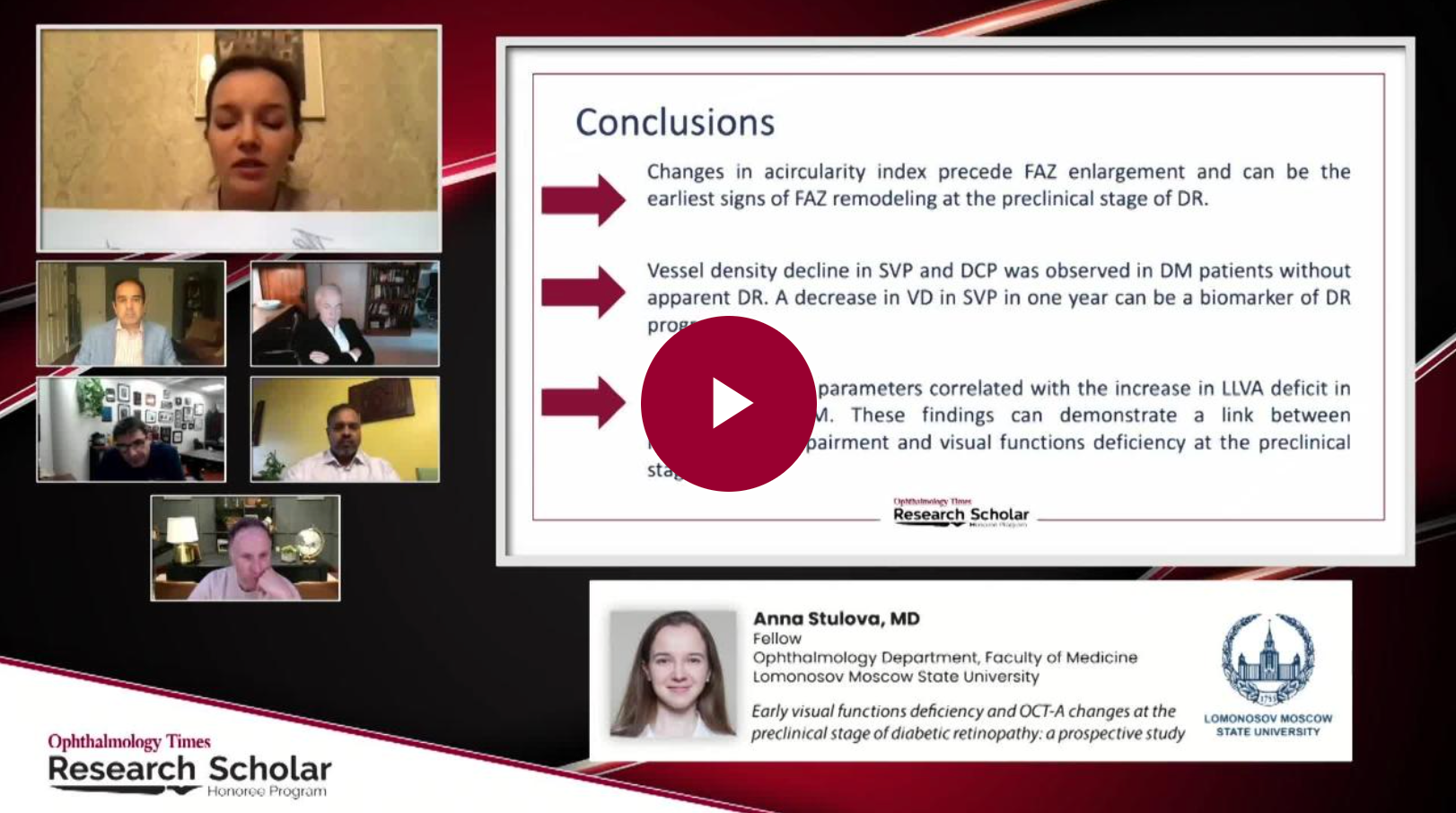
Conclusions
Our OCTA findings suggest changes in acircularity index precede FAZ enlargmement and can be the earliest signs of FAZ remodeling at the preclinical stage of DR.
We also see changes in foveal avascular zone circularity and parafoveal vessel density in SVP and DCP in patients with T1DM with no apparent DR.
The increase in LLVA deficit and its correlation with OCTA parameters demonstrate a link between retinal neurodegeneration and microvascular impairment at the preclinical stage of DR.
Further research will be done to assess these findings and evaluate their prognostic value.
OCT-A is popular in ophthalmology and will help manage patients with high risk. They will need 1-year examinations., which is the standard, and even more frequently.
See more submissions from the 2020 Research Scholar Honoree Program
About the author
Anna N. Stulova, MD
E: anna_stulova@mail.ru
Stulova is affiliated with the Ophthalmology Department of Lomonosov Moscow State University in Russia.